Portfolio Dog & cat

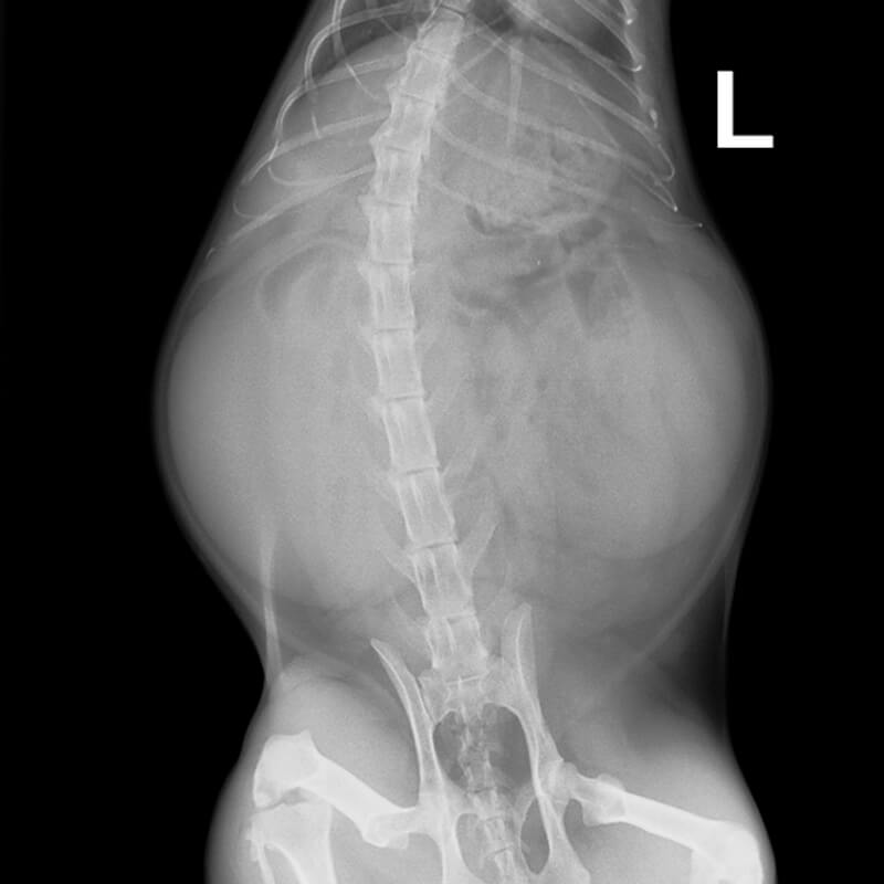
Rottweiler, Lexi, 4 years old

Generalized facial swelling, more pronounced on the left side; deformation of the jaw; no improvement with treatment (antibiotics, NSAIDs, glucocorticoids); PU/PD; azotemia

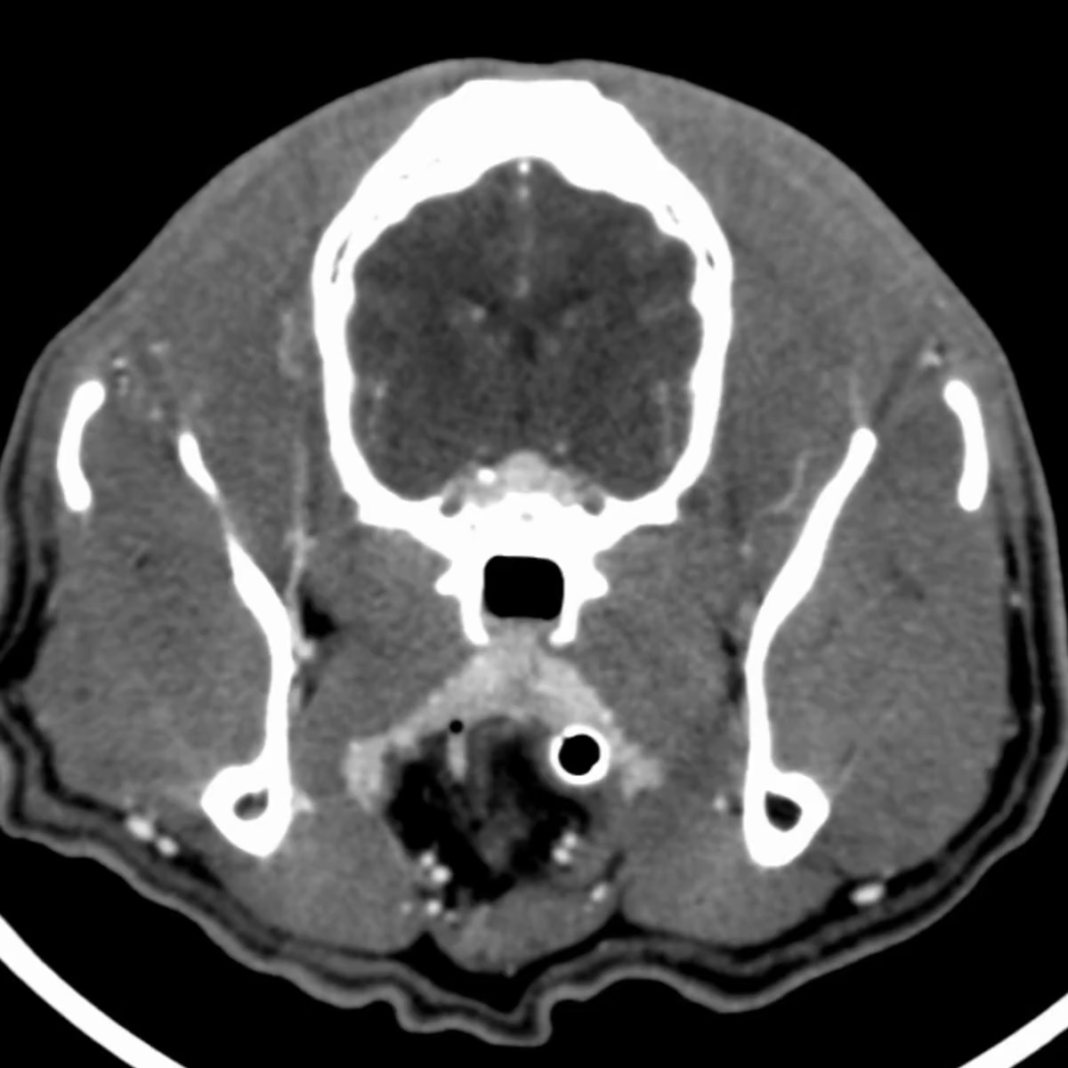
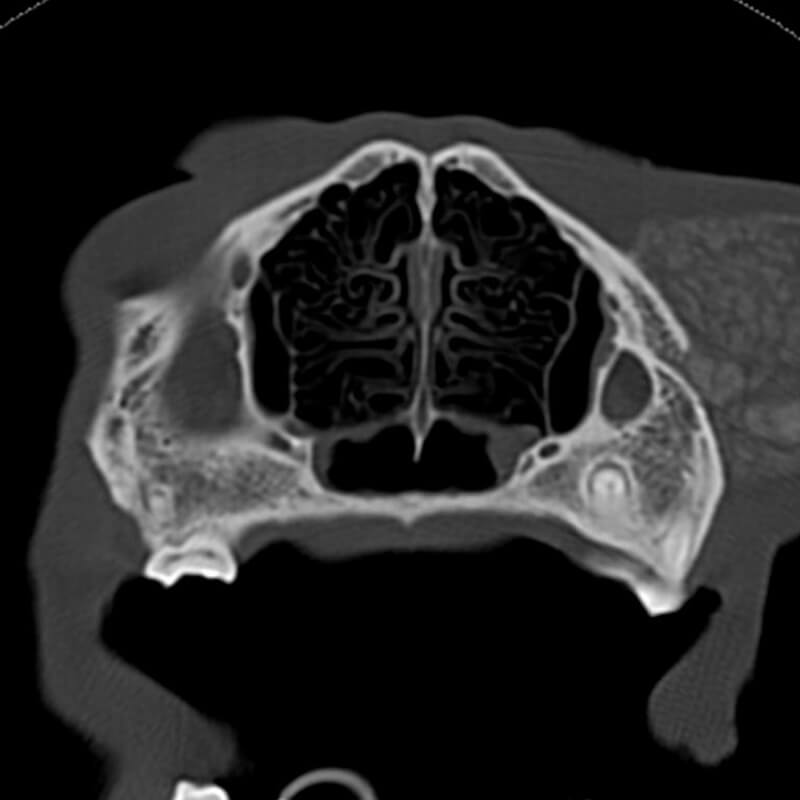
CT scan of the head, native sequence, bone reconstruction

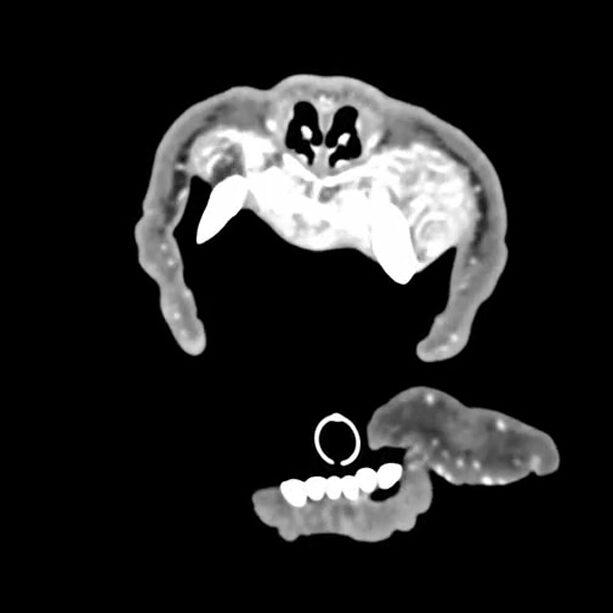
CT scan of the head, sequence after contrast administration, soft tissue reconstruction

Diagnosis

CT-Findings

The CT scan of the head shows a complete loss of the bony structure of the maxilla and a nearly complete loss of the bony structure of the mandible. Extensive areas of osteolysis with loss of the cortical bone are visible. Both bones are highly fibrous and distended, containing hyperattenuating onion-skin-like or halo-like material that takes up contrast medium in a highly heterogeneous manner. The remaining teeth are “floating” and have no connection to bony structures.

All other skull bones, including those of both the splanchnocranium and the neurocranium, show severe osteopenia with thinning of the cortical bone, loss of the trabecular structure, and areas of osteolysis.

Note

In addition to the bony changes, prominent epithelial bodies are visible on both sides of the thyroid gland. The complete CT scan also includes images of the thorax and abdomen. The kidneys appear markedly atrophic on both sides, with an irregular, nodular surface and reduced contrast enhancement. The findings in the kidneys are consistent with bilateral atrophic kidneys (nephrocirrhosis).

CT-Diagnosis

  • Severe fibrous osteodystrophy (rubber jaw syndrome)
  • Severe osteopenia of the facial skeleton and neurocranium
  • Generalized enlargement of the parathyroid glands
  • Bilateral kidney atrophy (nephrocirrhosis)

Discussion

The CT scan findings of “Lexi’s” head show the consequences of secondary renal hyperparathyroidism, including advanced generalized osteopenia of the skull bones and what is known as fibrous osteodystrophy of the jaw bones (rubber jaw osteodystrophy). Fibrous osteodystrophy is a metabolic bone disease in which increased bone resorption and its replacement by collagenous connective tissue lead to reduced bone density (osteopenia).

The cause is a prolonged elevation of parathyroid hormone levels in the blood, which results in the following pleiotropic effects:

Effect on the bones Effects on the kidneys Effect on the intestines
  • Indirect stimulation of osteoclasts
  • Release of calcium and phosphate from bone
  • increased calcitriol synthesis
  • increased renal calcium reabsorption
  • increased calcium absorption in the intestine
  • Stimulation of renal phosphate excretion

This hyperparathyroidism can be primary (endocrine-active hyperplasia, adenomas, or adenocarcinomas of the parathyroid gland), secondary to renal disease (resulting from chronic kidney disease), or tertiary (continuously high PTH secretion in the course of secondary hyperparathyroidism that is no longer subject to normal regulatory mechanisms).

In Lexi’s case, the atrophic kidneys and azotemia indicate the presence of chronic renal insufficiency. Lexi’s enlarged parathyroid glands respond to the resulting hyperphosphatemia and hypocalcemia by overproducing parathyroid hormone, which in turn causes a severe disturbance in calcium homeostasis (renal hyperparathyroidism).

References

  • Baumgärtner, W., Ulrich, R. (2020). Osteodystrophia fibrosa. In: Baumgärtner W, Gruber A, Hrsg. Spezielle Pathologie für die Tiermedizin (2., aktualisierte Auflage). Stuttgart: Thieme.
  • Brachthäuser, L., Pingen, C. H., Hecht, W., Reinacher, M. (2013). Rubber jaw in a Weimaraner dog due to juvenile nephropathy. A case without evidence for genetic involvement. Tierärztliche Praxis Ausgabe K: Kleintiere / Heimtiere, 41(3), 198–202.
  • Grünberg, W., Ramirez, A. (2020). Fibrous Osteodystrophy in Animals. MSD Veterinary Manual. https://www.msdvetmanual.com/musculoskeletal-system/dystrophies-associated-with-calcium-phosphorus-and-vitamin-d/fibrous-osteodystrophy-in-animals
  • Kyle, M. G., Davis, G. B., Thompson, K. G. (1985). Renal osteodystrophy with facial hyperostosis and ‘rubber jaw’ in an adult dog. New Zealand Veterinary Journal, 33(7), 118–120.
  • Waller, S. B., Correia Canuto, F. J., Correia Costa, P. P., Louzada Dias Cavalcanti, E. A. N., de Oliveira Cavalcanti, G. A., Vasconcelos, M., & Blum Cleff, M. (2019). Maxillomandibular Deformity in a Canine with Fibrous Osteodystrophy Secondary to Chronic Kidney Disease. Acta Scientiae Veterinariae, 47 (Suppl 1).
Read More

French Bulldog, Freya, 10 years

Hematochezia and suspected rectal neoplasia
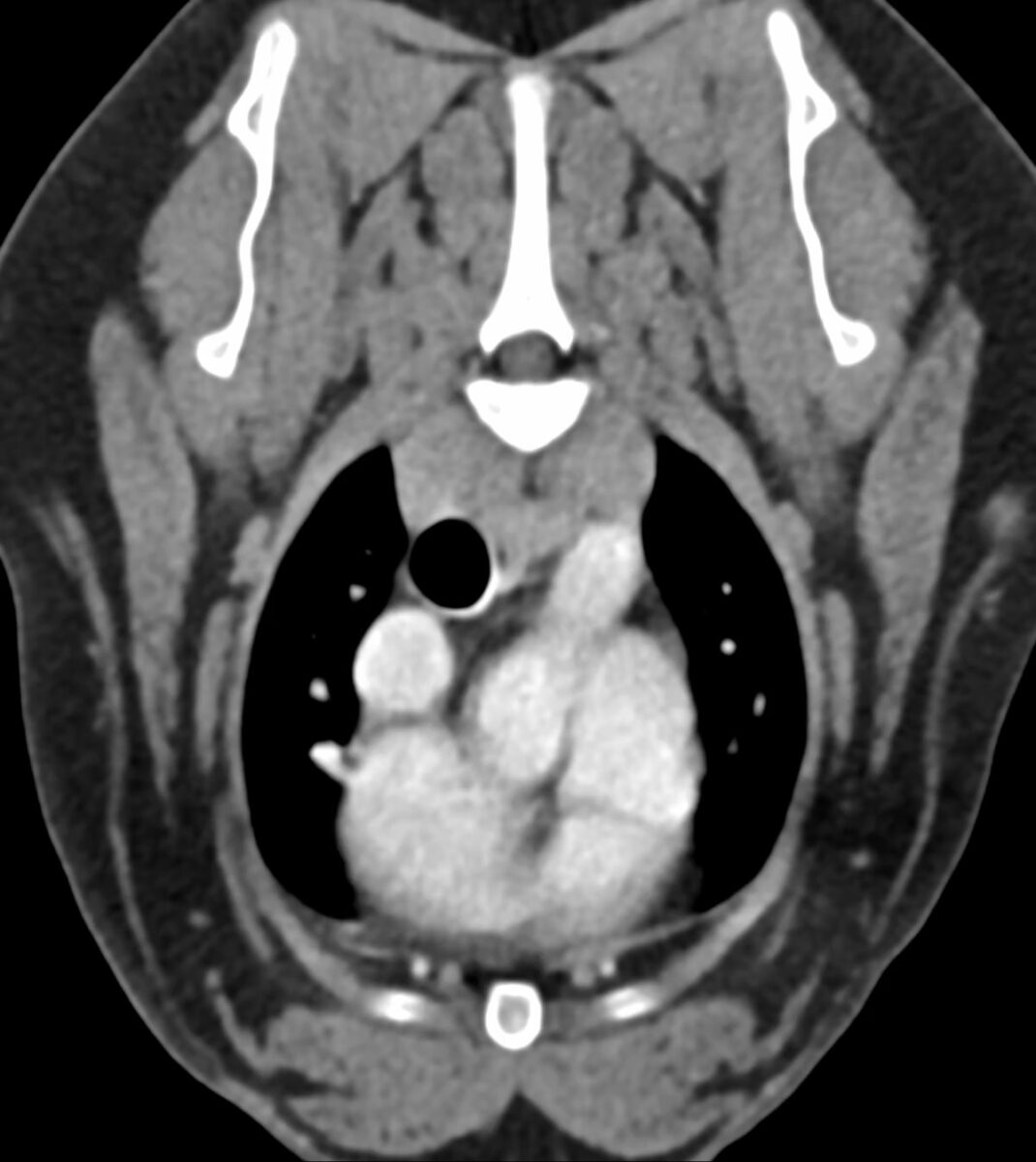
Heart base mass noted on CT

CT dated Mai 2025, with a mass noted at the base of the heart, which was an incidental finding and asymptomatic at the time of imaging.

Diagnosis

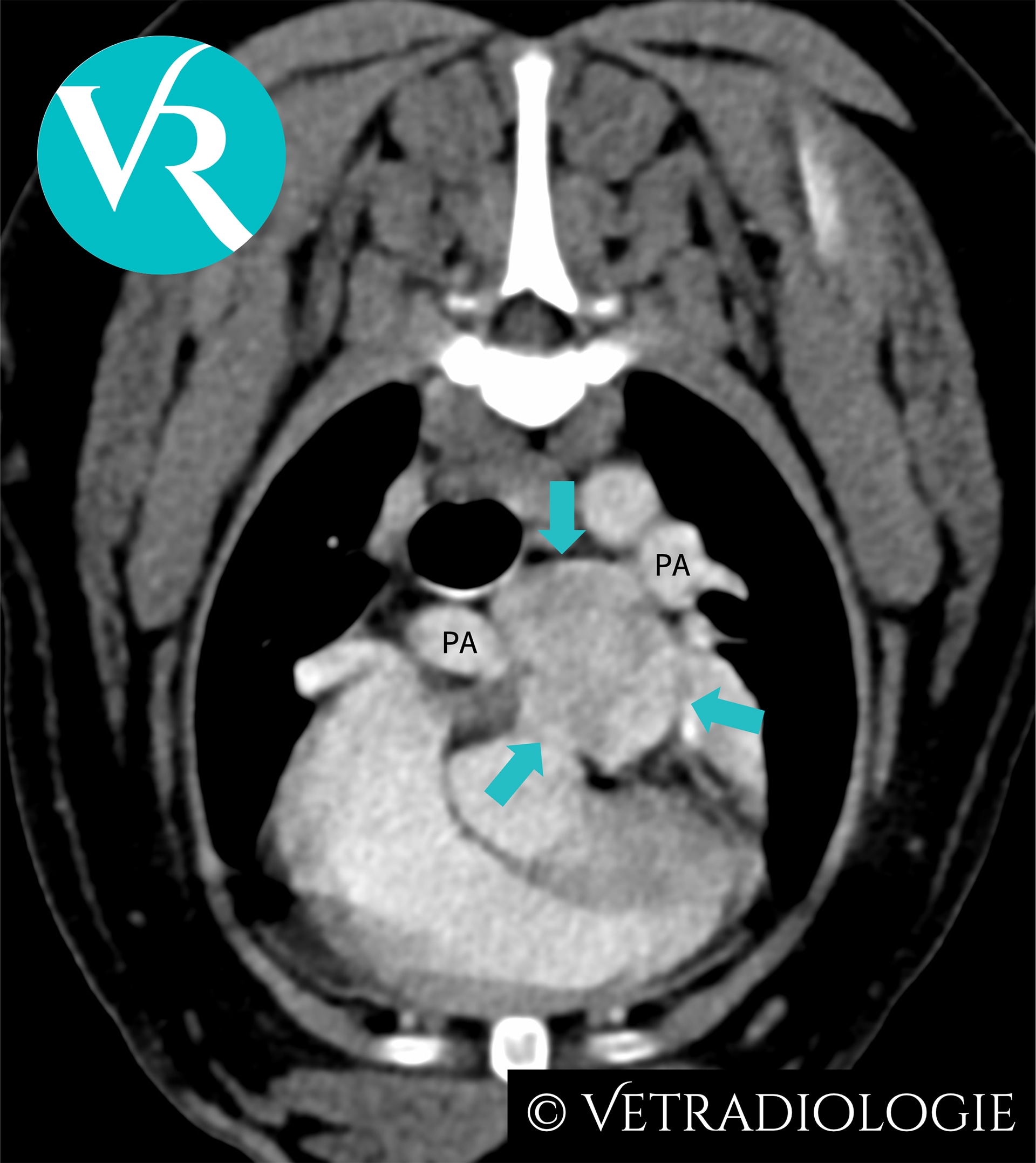
Freya’s CT Study: Note the well circumscribed, ovoid, heterogeneously contrast enhancing soft tissue mass (arrows) at the level of the main pulmonary arterial bifurcation.

CT-Findings

There is a well circumscribed, ovoid, hypervascular soft tissue mass (approximately 2.6cm in maximum diameter) located at the level of the main pulmonary arterial bifurcation, exhibiting heterogeneous contrast enhancement.

CT-Diagnosis

  • Heart base tumor

Discussion

Location and morphology are characteristic for a chemodectoma originating from the pulmonary glomus. A chemodectoma is a type of neuroendocrine tumor (NET) arising from chemoreceptor cells at the main pulmonary trunk. These chemoreceptors are organized into glomera and functionally assigned to the paraganglia. Tumors of these circulatory regulatory structures are referred to as chemodectomas or paragangliomas, and are reported more commonly in dogs than in cats.

Chemodectomas are typically benign, non-functional, and exhibit a low metastatic rate, although they may demonstrate locally invasive growth. Due to their slow growth, they frequently remain asymptomatic and—as in Freya—are often incidental findings. However, they may lead to clinical signs such as syncope, exercise intolerance, or sudden cardiac death.

In addition to the pulmonary glomus, chemodectomas/paragangliomas may also arise from other glomera such as from the aortic body glomus at the aortic root or carotid glomus at the bifurcation of the carotid artery.

The chemoreceptors of the glomera respond physiologically to reduced partial oxygen pressure and decreased blood pH. Chronic stimulation due to hypoxemia and respiratory acidosis, such as occurs in the context of the brachycephalic airway syndrome, has been discussed as a possible cause of the increased prevalence of chemodectomas in brachycephalic dogs

References

  • Buscaglia NA, Johnson PJ. What Is Your Diagnosis? J Am Vet Med Assoc. 2019;254(4):467-469. doi:10.2460/javma.254.4.467.
  • Crawford-Jennings MI, Chavez LD, Loessberg ER, Carvallo-Chaigneau FR. Aortic body tumor with intracardiac metastasis in a dog. J Vet Diagn Invest. 2025 Mar;37(2):345-348. doi: 10.1177/10406387241304438.
  • Fife W, Mattoon J, Drost WT, Groppe D, Wellman M. Imaging features of a presumed carotid body tumor in a dog. Vet Radiol Ultrasound. 2003;44(3):322-325. doi:10.1111/j.1740-8261.2003.tb00463.
  • Kromhout K, Gielen I, De Cock HE, et al. Magnetic resonance and computed tomography imaging of a carotid body tumor in a dog. Acta Vet Scand. 2012;54:24. doi:10.1186/1751-0147-54-24.
  • Mai W, Seiler GS, Lindl-Bylicki BJ, et al. CT and MRI features of carotid body paragangliomas in 16 dogs. Vet Radiol Ultrasound. 2015;56:374-383. doi:10.1111/vru.12251.
  • Obradovich JE, Withrow SJ, Powers BE, Walshaw R. Carotid body tumors in the dog. Eleven cases (1978–1988). J Vet Intern Med. 1992;6(2):96-101. doi:10.1111/j.1939-1676.1992.tb03158.
  • Wess G. Kardiale und perikardiale Tumoren. In: Kessler M, Hrsg. Kleintieronkologie. 4., vollständig überarbeitete Auflage. Stuttgart: Thieme; 2022:600f.
Read More

French Bulldog, Sammy, 9 years

Head tilt, right-sided Horner’s syndrome, right-sided facial discomfort

CT dated September 2021. Presented with 10d history of cough, dyspnea and nasal discharge. Acute right-sided facial paralysis and lethargy.

Diagnosis

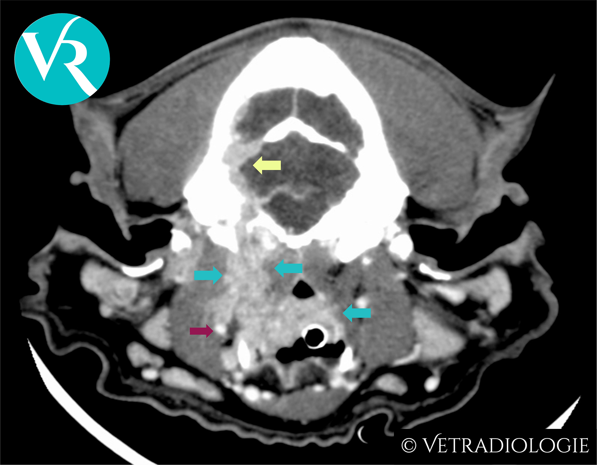
Sammy’s CT-Study: Infiltrative soft tissue mass (turquoise arrows) exhibiting marked heterogeneous contrast enhancement and invasion of the cranial vault (yellow arrow) along the plane of the right internal carotid artery (red arrow).

CT-Findings

Right-sided retropharyngeal soft tissue mass located medial to the right mandibular salivary gland in the region of the common carotid arterial bifurcation, with craniodorsal mass extension towards the cranial vault. There is associated invasion of the digastric and medial pterygoid muscles, and osteolysis of the right temporal bone (including petrous, tympanic and squamous parts). There is extension of the lesion into the caudal fossa via moth-eaten and permeative osteolysis of the squamous part of the right temporal bone, with the intracranial mass component reaching and conforming to the tentorium cerebelli ventrally.

Caudally, the lesion extends along the right common carotid artery to the approximate level of the thyroid gland.
The lesion exhibits marked, heterogeneous contrast enhancement.

CT-Diagnosis

  • Aggressive, invasive soft tissue mass originating near the bifurcation of the right common carotid artery.

Discussion

The imaging features of the mass are consistent with a carotid body tumor/glomus caroticum tumor. This subtype of paraganglioma belongs to the group of neuroendocrine tumors (NETs).

Brachycephalic dog breeds are predisposed.

Carotid body tumors arise from the neuroendocrine chemoreceptors of the carotid glomus, located at the bifurcation of the common carotid artery. Similar clusters of chemoreceptors (glomera) exist in the wall of the aorta (aortic body glomus) and the pulmonary trunk (pulmonary glomus), where tumor development (chemodectomas, glomus aorticum tumors) has likewise been described.

As circulatory regulatory structures, the chemoreceptors of the glomera respond to reduced oxygen partial pressure and to changes in blood pH.

Chronic hypoxia and respiratory acidosis associated with brachycephalic obstructive airway syndrome (BOAS) lead to persistent stimulation of these chemoreceptors. It is assumed that this—either alone or in combination with a genetic component—accounts for the described breed predisposition to the development of carotid body tumors and chemodectomas.

The clinical presentation varies depending on tumor size and the extent of involvement of adjacent structures. Potential symptoms include regional discomfort, head tilt, coughing, dyspnea, Horner’s syndrome, or dysphagia.

On CT, carotid body tumors appear as highly vascularized, locally invasive masses with marked, heterogeneous contrast enhancement.

References

  • Buscaglia NA, Johnson PJ. What Is Your Diagnosis? J Am Vet Med Assoc. 2019;254(4):467-469. doi:10.2460/javma.254.4.467.
  • Crawford-Jennings MI, Chavez LD, Loessberg ER, Carvallo-Chaigneau FR. Aortic body tumor with intracardiac metastasis in a dog. J Vet Diagn Invest. 2025 Mar;37(2):345-348. doi: 10.1177/10406387241304438.
  • Fife W, Mattoon J, Drost WT, Groppe D, Wellman M. Imaging features of a presumed carotid body tumor in a dog. Vet Radiol Ultrasound. 2003;44(3):322-325. doi:10.1111/j.1740-8261.2003.tb00463.
  • Kromhout K, Gielen I, De Cock HE, et al. Magnetic resonance and computed tomography imaging of a carotid body tumor in a dog. Acta Vet Scand. 2012;54:24. doi:10.1186/1751-0147-54-24.
  • Mai W, Seiler GS, Lindl-Bylicki BJ, et al. CT and MRI features of carotid body paragangliomas in 16 dogs. Vet Radiol Ultrasound. 2015;56:374-383. doi:10.1111/vru.12251.
  • Obradovich JE, Withrow SJ, Powers BE, Walshaw R. Carotid body tumors in the dog. Eleven cases (1978–1988). J Vet Intern Med. 1992;6(2):96-101. doi:10.1111/j.1939-1676.1992.tb03158.
  • Wess G. Kardiale und perikardiale Tumoren. In: Kessler M, Hrsg. Kleintieronkologie. 4., vollständig überarbeitete Auflage. Stuttgart: Thieme; 2022:600f.
Read More

DSH cat Napoleon, 5 years

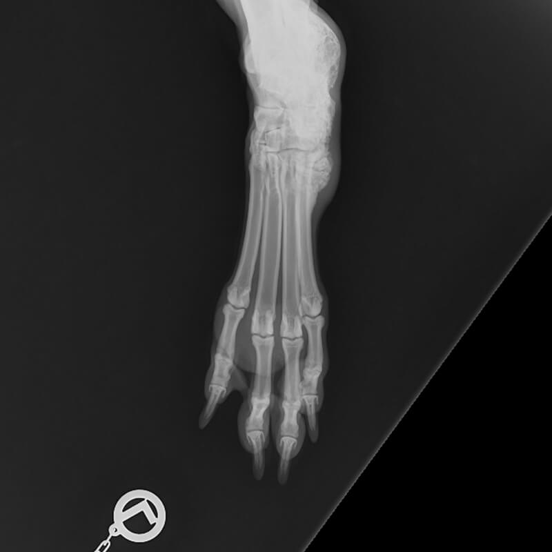
R forelimb lameness

With kind permission of the small animal clinic Frankenthal

Diagnosis

Description

A 6 cm long and 4 x 4 mm wide bone fragment is located on the caudo-lateral aspect of the right ulna. The bone fragment begins 2 cm distal to the Tuber olecrani, tapers and ends in a pointed tip. The fragment is sclerotic when compared to the adjacent cortex and is surrounded by a radiolucent rim. A severe brush border periosteal reaction extends from the olecranon along the ulna to end distal of the fragment. The periosteal new bone appears more lucent when compared to the cortical opacity.

An isolated, elongated, smoothly marginated mineral opacity is evident cranial to the humeral condyles.

Radiographic diagnoses

  • Sequestrum right ulna with severe surrounding periostitis
  • Small bony fragment cranial to humeral condyles

Discussion

The changes are suggestive of a traumatic insult which has led to sequestrum formation of the ulna. The small bony fragment cranial to the humeral condyles can also be traumatic in origin and represent for example a chip fracture of the radial head. Differential diagnosis is dystrophic mineralisation of a tendon.

Read More

Labrador Retriever Bella, 9 years

Progressive swallowing problems, especially of larger items

Post-contrast sequence

Pre-contrast sequence

With kind permission Dr. G. Viefhues, Small Animal Clinic Ahlen

Diagnosis

Description

The atlanto-occipital and atlanto-axial junctions are surrounded by a cavitating, space occupying lesion which is hypoattenuating to the surrounding soft tissue. The density of the individual cavities is 11-13 HU (density of the neighboring muscles is approx. 80 HU). Another space occupying lesion is located ventral to the M. longus capitis and appears separate from the other masses. It is approx. 3.8cm long, 2.5cm high, 4.1cm wide and results in severe displacement of the pharyngeal roof so that the caudal nasopharynx is completely obliterated. The occipital and temporal bones as well as the atlas (C1) and cranial aspect of the axis (C2) including the dens show a large number of well circumscribed, lytic defects. The space occupying lesion extends into the right caudal cranial fossa which leads to mild displacement and compression of the cerebellar vermis. Brain stem and spinal cord at C1 and C2 are also compressed; mostly from ventral and the sides. Severe, heterogenous and foamy contrast enhancement of the compressing structures in the spinal canal is evident. The space occupying lesions do not contrast enhance.

CT diagnosis

  • Soft tissue neoplasia atlanto-occipital and atlanto-axial junction with bone destruction and compression of cerebellum, pons and spinal cord

Discussion

The changes are suggestive of a myxosarcoma. Differential diagnoses include all other soft tissue tumours; however, the extreme hypodensity of all masses is highly suggestive of a myxosarcoma. The tumor is most likely arising from the atlanto-occipital and atlanto-axial joints, which are continuous which each other. The described masses are therefore most likely connected and part of one tumor mass.

Outcome

Bella was euthanized. The histopathological report confirmed the diagnosis of myxosarcoma.

Read More

DSH cat Lucy, 8 years

Mild cough

With kind permission Dipl.med.vet. TA Hennig

Diagnosis

Description

Body condition score (BCS) 5-6/9. The skeletal structures are physiological. The caudo-ventral liver lobe is rounded and extends just beyond the costal arch. A moderate amount of air is present in the stomach. The gastric axis is parallel to the ribs. A soft tissue structure with a triangular cranial outline is evident ventro-caudal to the stomach; its caudal component is not included in the view (tail of spleen). As far as included in the images, the retroperitoneal and peritoneal detail is good.

The thoracic volume is physiological and the contour of the diaphragm is relatively straight.

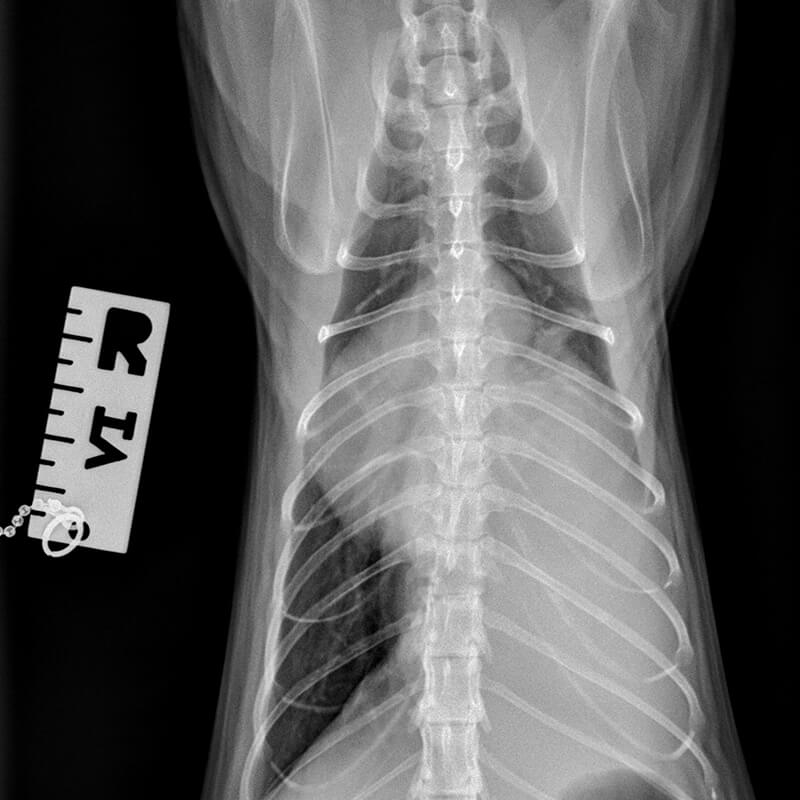
The left hemithorax is filled with a soft tissue opaque mass extending from rib 6 to the diaphragm. The contour of the mass is slightly rounded on the cranial, ventral and medial aspects. Silhouette signs exists between mass, heart and left diaphragm. Carina and cardiac shadow have been displaced ventrally and to the right by the mass; the left main stem bronchus is also displaced ventrally. The lobar bronchus for the diaphragmatic lobe (right caudal lobe) can only be appreciated just caudal to the carina.

The cardiac silhouette shows no obvious changes. The pulmonary vessels also appear physiological.

Radiological diagnoses

  • Space occupying lesion left caudal thorax
  • Aerophagia
  • Slightly rounded caudo-ventral liver lobe

Discussion

The most likely cause for the mass is a tumour in the left caudal lobe. Differential diagnoses include granuloma and abscess.

A diaphragmatic rupture can be ruled out due to a physiological position of the stomach. An extra-pleural mass is also unlikely due to the described radiographic appearance.

The aerophagia is most likely stress related however, small intestinal loops are not included in the views thus chronicity cannot be ruled out entirely. Aerophagia due to dyspnea is a differential diagnosis.

The rounded appearance of the ventral liver lobe is most likely due to the projection of the gall bladder over the hepatic contour which is a physiological occurrence in the feline species. The prominence can be the result of prolonged inappetence but outflow obstruction is a differential diagnosis.

Outcome

Histo-pathology revealed a carcinomatous process in the lung; most likely a broncho-alveolar carcinoma. Adenocarcinoma is a differential diagnosis.

Read More

DSH cat Charly, 12 years

Palpable abdominal masses

With kind permission of Drs. David and Krützfeldt, Small Animal Clinic Frankenthal

Diagnosis

Description

The radiographs are of diagnostic quality; both lateral and VD views are slightly rotated.

Body condition score (BCS) 5/9.

The disc spaces T12-L3 are narrow and the endplates are deformed.

The abdominal detail is good. Both renal shadows are severely enlarged. The right renal shadow is 5.5 and the left 4 x the length of the vertebral body of L2 (normal: 1.9 – 3.2x the length of L2 vertebral body). The renal contours are smooth. Their shape is oval with loss of the characteristic indentation at the level of the renal hilus. The opacity is that of soft tissue. The renal enlargement causes a severe mass effect with displacement of the adjacent organs. The stomach contains a moderate amount of food and is cranially displaced; its caudal contour is concave. The intestinal tract is severely displaced ventrally. The small intestine is mostly empty and the colon contains formed faeces and gas. The other abdominal structures appear physiological.

Radiological diagnoses

  • Severe bilateral renomegaly
  • Degenerative disc disease T12-L3

Discussion

Differential diagnoses in case of bilateral renomegaly with smooth surfaces, physiological shape and opacity include perirenal pseudocyst, diffuse, infiltrative neoplasia such as lymphoma and hydronephrosis.

Outcome

On ultrasound a large amount of anechoic, subcapsular fluid surrounded both kidneys. This is compatible with perirenal pseudocyst (bilateral).

Perirenal pseudocyst describes a peri-renal, mostly subcapsular, fluid accumulation which usually represents a transudate. The cause is unknown. Often chronic renal disease is present though a causative effect has yet to be proven.

US image: with kind permission of the Small Animal Clinic Frankenthal

Read More

Staffordshire terrier Gizmo, 10 years

Bilateral toe dragging hind limbs, progressive paraparesis

T2 weighted

T2 weighted

T1 weighted without contrast

T1 weighted with i.v. contrast

With kind permission Dr. O. Lautersack, Tierärztliche Klinik für Chirurgie Ettlingen

Diagnosis

MRI description

In the T2 weighted images all included discs show a varying degree of signal loss of the nucleus pulposus. A moderate to severe degree of spondylosis is located at the TL junction and the first three lumbar vertebrae. An approx. 2cm long mass is present to the L of the spinal cord at the level of L2. At the level of the intervertebral foramen L1/2 it causes compression and displacement of the spinal cord to the right. Centrally over L2 the spinal cord is completely abnormal. The transverse images do not indicate if the spinal cord is completely infiltrated or merely severely displaced and compressed. On the T2 weighted dorsal image a “Golf-T sign”, which represents widening of the subarachnoid space, is present cranial and caudal to the lesion. In the T2 weighted images the mass is of heterogeneous hyperintensity, in the T1 weighted images it is hypointense with strong enhancement. A thin structure without contrast enhancement is evident ventral and to the R of L2; its signal intensity is that of the normal spinal cord.

On the sagittal views only part of the LS junction is included. The LS disc appears to protrude moderately into the spinal canal, replacing the ventral epidural fat so that it comes into contact with the cauda equine.

MRI diagnoses

  • Intradural, mostly extramedullary and partially intramedullary mass level with L2
  • Generalised disc degeneration
  • Spondylosis TL junction and cranial L-spine
  • Cauda equine compression secondary to disc protrusion

Discussion

The list of differential diagnoses includes peripheral nerve sheath tumor, lymphoma, malignant histiocytic sarcoma and meningioma. However, the appearance is not typical for the last three tumor types. Localisation and behavior of the mass also fit the diagnosis of neuroblastoma; however, clinical signs generally begin to show between the first and third year of life. In case the tumor is slow growing, the onset of clinical signs can be delayed until later in life.

Outcome

Photo after durotomy.
With kind permission Dr. O. Lautersack.

Gizmo’s owner elected surgery.

During surgery the tumor was intradurally located and had a greasy, friable consistency. Only a partial resection was possible.

The histological diagnosis was atypical meningioma (Grade II WHO).

Two weeks post operatively “Gizmo” had improved significantly and could walk without signs of paraparesis.

Read More

GSD Tyson, 2 years

Lameness of both hind limbs with swelling around both tarsal joints

The radiographic changes affect both tarsal joints. The images shown here are of the L tarsus.

With kind permission Drs. Kessler, Kosfeld, Tassani-Prell, Bessmann, Rupp, Delfs, Schmohl, von Klopmann Tierklinik Hofheim.

Diagnosis

Description

A large amount of bridging new bone is present on the dorsal, plantar and lateral aspects of tibia and especially at the tibio-tarsal joint. The surface of the new bone formation is smoot and the opacity heterogeneous. The bone opacity of the talus is reduced.

Radiological diagnosis

  • Bony mass around tarsus with ankyloses of tibiotarsal- and intertarsal joints
  • Severe osteopenia talus

Discussion

The list of differential diagnoses includes multiple cartilaginous exostoses and changes associated with diffuse, idiopathic hyperostosis (DISH). The bilateral symmetrical nature of the changes would be unusual for a traumatic cause. The radiological findings are not compatible with enthesiophytosis or degenerative joint disease.

Due to Wolff’s law, a lack of bone loading results in bone loss; thus the osteopenia of the talus is the result of disuse due to ankylosis. Due to the traction of the Achilles tendon, which causes some loading and thus stimulation of bone turnover, the calcaneus is affected to a lesser degree. Assessment of the degree of osteopenia of the central tarsal and 1st row of tarsal bones is impossible due to superimpositioning. A small degree of osteopenia affecting the distal tarsal bones could be present and could be explained by a small degree of retained movement in the tarso-metatarsal joints.

Outcome

Due to the absence of vertebral abnormalities such as ossification of the ventral longitudinal ligament, as would be expected in DISH, it is likely that “Tyson” is suffering from a form of multiple cartilaginous exostoses.

Read More

Small Münsterländer Samson, 7 years

For 4 weeks: swelling lateral to the L eye, no improvement on antibiotics and pain killers: trauma cannot be ruled out.

With kind permission Dr. G. Viefhues, Small Animal Clinic Ahlen

Diagnosis

Description

An approx. 3cm large, mineral dense mass with a granular appearance extends from the zygomatic process of the left maxilla to the zygomatic arch. The mass protrudes slightly into the orbital cavity and causes mild deformity of the globe. The underlying cortex has a slightly roughened surface and shows small, fine osteolytic regions. An extension of the mass into the bone marrow is not evident.

CT diagnosis

  • Bony mass left zygomatic arch

Discussion

The appearance of the tumor is typical for a multilobular tumor of the bone. Other names are osteochonrosarcoma and chondrosarcoma rodens.

Read More
WordPress Cookie Notice by Real Cookie Banner