Portfolio Dog & cat

Irish Wolfhound Bailey, 1 year

Initially left sided, purulent nasal discharge, later bilateral nasal discharge

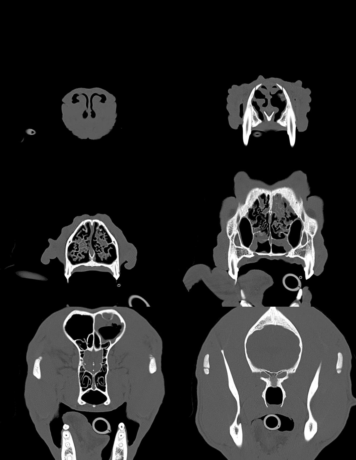
CT images from July 2014. At that time unilateral nasal discharge was present.
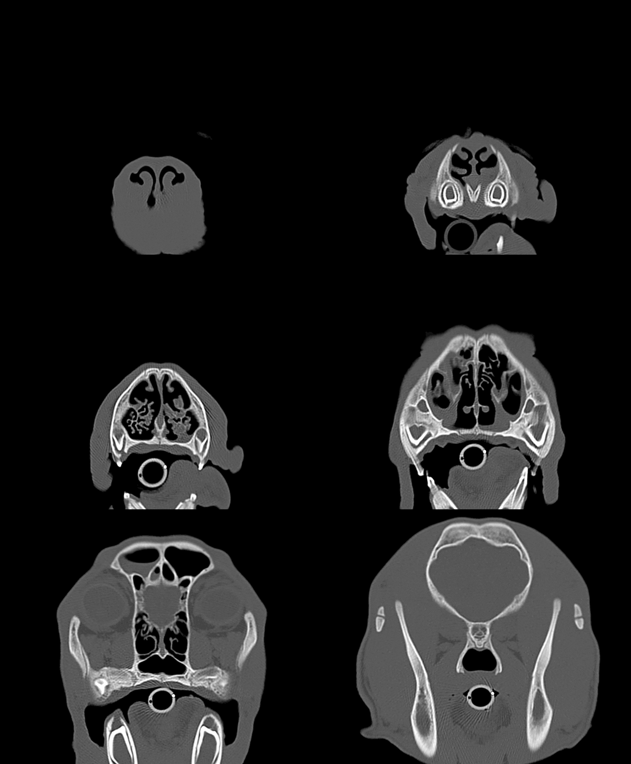
CT images from March 2015. This time bilateral nasal discharge was present.
Images courtesy of Kleintierklinik Greven. Dres. Böhmer, Cordes, Möller and Wienker

Diagnosis

Description

Severe reduction in size and number of the turbinates in both nasal cavities. Between the remaining turbinates a mild to moderate amount of soft tissue dense material is present. Within the left frontal sinus soft tissue dense material with a fluid level is present (images flipped horizontally, patient was placed in dorsal recumbency during image acquisition). The mucosal lining of the left frontal sinus is thickened. Findings are almost identical in both CT studies.

Radiographic diagnoses

  • Bilateral reduction in number and size of nasal turbinates
  • Soft tissue dense material between the turbinates
  • Sinusitis with fluid accumulation left frontal sinus

Discussion

Changes are compatible with rhinitis- bronchopneumonia syndrome of the Irish Wolfhound. The etiology is unclear. Immune mediated or primary ciliary defects are discussed. A hereditary background is assumed. Affected dogs show transient or persistent mucoid to mucopurulent nasal discharge of variable degree from birth . Concurrent bronchopneumonia can occur (most likely due to the ciliar dysfunction leading to an increased susceptibility for infection).

No progression of the changes is seen over the one year period. No granuloma is visible in the frontal sinus. The paranasal bones are unremarkable, showing no permeative osteolysis. Therefore a fungal rhinitis is less likely. In addition the dog is very young making a fungal rhinitis unlikely as well.

Recommendations

None.

Normal CT study nasal cavity

A reduction in the number of turbinates is not always related to a destructive rhinitis. Consider congenital hypoplasia predisposing the nasal cavity to infection, especially in young dogs.

Read More

Crossbreed dog Lucky, 9 years

Swallowing problems, respiratory distress

Images courtesy of Tierklinik Hofheim. Dres Kessler, Kosfeld, Tassani-Prell, Bessmann, Rupp, Delfs, Schmohl, von Klopmann

Diagnosis

Description

Good body condition. Inspiratory radiograph.

Mild new bone formation visible ventral to T4/5 and T12/13 (spondylosis). Small osteophyte visible at the infraglenoid tubercles of one scapula. Abdominal detail within normal limits.

There is mild convex bulging of the ventral border of the cranial mediastinum. Overall the opacity at the level of the cranial mediastinum appears increased. The trachea runs almost parallel to the thoracic spine with a mild dorsal buldging at the level of the 3rd to 4th intercostal space. Dorsal to the 2nd sternebra a poorly delineated soft tissue opaque structure is visible.

The lung shows an overall, severely increased opacity. Between the cardiac silhouette and the diaphragm it is of almost soft tissue opacity, however the diaphragmatic outline can still be identified. On close inspection also fine white lines can also be seen in the affected areas; they represent vessels. The remaining lung shows more reticular changes. Throughout the entire lung field multiple, soft tissue nodules are visible.

Radiographic diagnoses

  • Severe interstitial pattern with multiple small soft tissue nodules (reticulonodular)
  • Convexity of the cranial mediastinum
  • Soft tissue dens structure dorsal to the 2nd sternebra
  • Spondylosis
  • Mild osteoarthrosis shoulder

Discussion

The lung changes are highly suspicious for a neoplasia (primary versus secondary).

The changes of the cranial mediastinum are suspicious for a cranial mediastinal mass. Differential diagnoses include enlarged cranial mediastinal lymphnodes, mass arising from the oesophagus (neoplasia, granuloma) as well as mediastinal haemorrhage, abcesses, granuloma or inflammation (for example due to perforation of the oesophagus, nocardiosis).

The soft tissue structure dorsal to the 2nd sternebra is most likely an enlarged sternal lymphnode.

The severe opacification caudodorsal to the cardiac silhouette can be due to superimposition of severely altered lung parenchyma. As only a single lateral radiograph is available a caudal mediastinal mass, for example arising from the oesophagus, a hematoma, an abcess or a granuloma cannot be excluded. A second radiograph would be required to assess this change further.

In combination with the lung changes a neoplasia with metastasis to the sternal and mediastinal lymphnodes appears the most likely differential diagnosis.

Recommendations

  • Left lateral and ventrodorsal radiograph
  • And/or Computed tomography of the lung

Outcome

Computed tomography of the thorax confirmed the severe interstitial lung pattern with multiple soft tissue nodules. The sternal as well as the cranial mediastinal lymphnodes were severely enlarged.

Caudal to the heart no other mass lesions were visible, therefore the radiographic changes are most likely due to superimposition of the lung changes.

Lucky had a carcinoma of the larynx with metastasis to the lung and lymphnodes.

A heavy interstitial lung pattern can mimic an alveolar lung pattern. Don’t get fooled by the first impression. On close inspection there are usually still some vessels visible.

Read More
WordPress Cookie Notice by Real Cookie Banner