Made easy
Our service for you. To look something up quickly, you have access to a free of charge, always available, simple guide to topics that are deemed difficult.
What is the most likely diagnosis for which lung pattern? Which part of the heart can be found where on the cardiac silhouette? Which course does a portosystemic shunt take?
Lung pattern
Who doesn’t know the feeling: you look at a chest radiograph see a bit of every lung pattern and end up with a diagnosis of bronchio-interstitial infiltrate with alveolar components. It is almost impossible to reach a clinical diagnosis from this. But what does a good radiological description of a lung pattern, from which the most likely clinical diagnosis can be derived, look like? Here you will find a step-by-step guide for the evaluation of the lung, at the end of which a list of possible differential diagnoses can be drawn up.
Diagnosis of cardiac diseases
Of course, no cardiac diagnosis can be made based on a radiograph alone. However, a radiograph is certainly able to point to more than just the diagnosis of “cardiomegaly” and “pulmonary oedema yes/no”. In some cases, an accurate assessment of an altered cardiac silhouette may narrow down the list of differential diagnoses to one or two probable ones. Take a look – and see how this works.
Portosystemic shunts
Which forms of congenital portosystemic shunt (PSS) can we distinguish? Is there an easy way to remember the classic shunt forms in dogs? Here is a guide to help you accurately diagnose the next portosystemic shunt patient. Sometimes the home straight is in fact a loop.
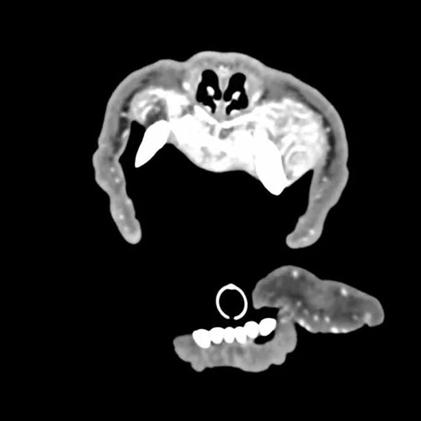
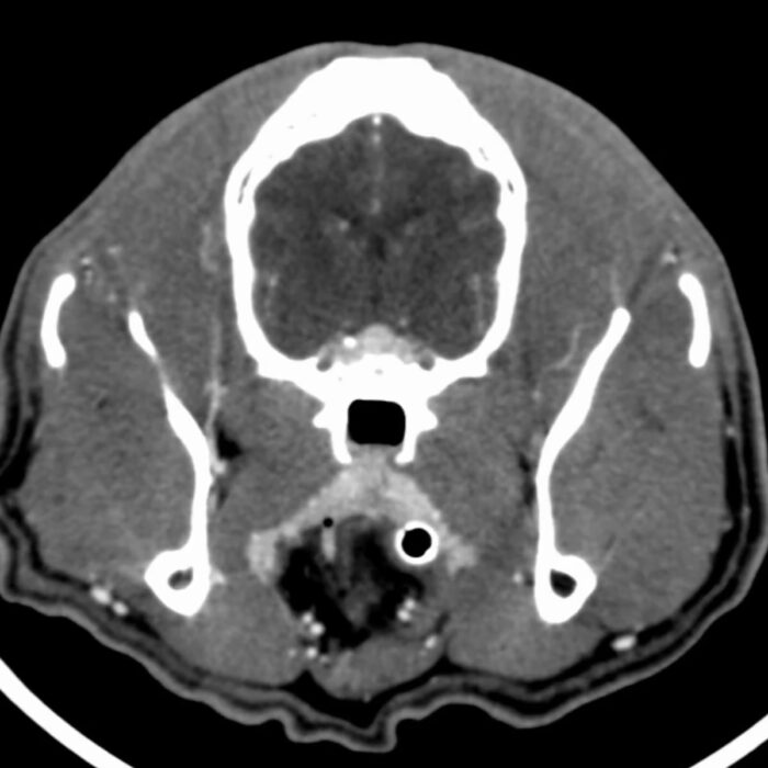
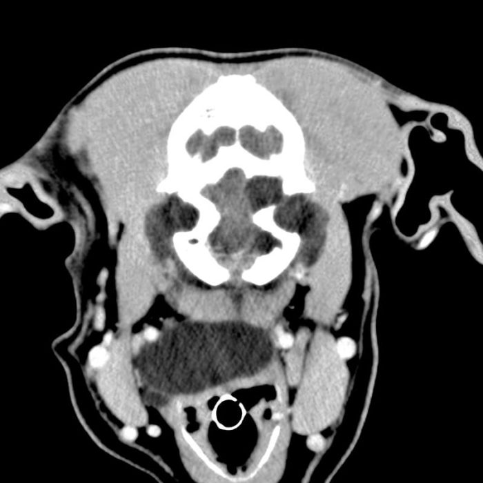
Browse through our case studies. Would you have known? Using MRI, CT, X-ray or ultrasound image material, you can make your own diagnosis and compare it with our findings.