Portfolio X-ray

Maltese dog Jimmy, 9 years

For last 6 months coughing after drinking

Images courtesy of Dr. G. Viefhues Tierärztliche Klinik Ahlen

Diagnosis

Description

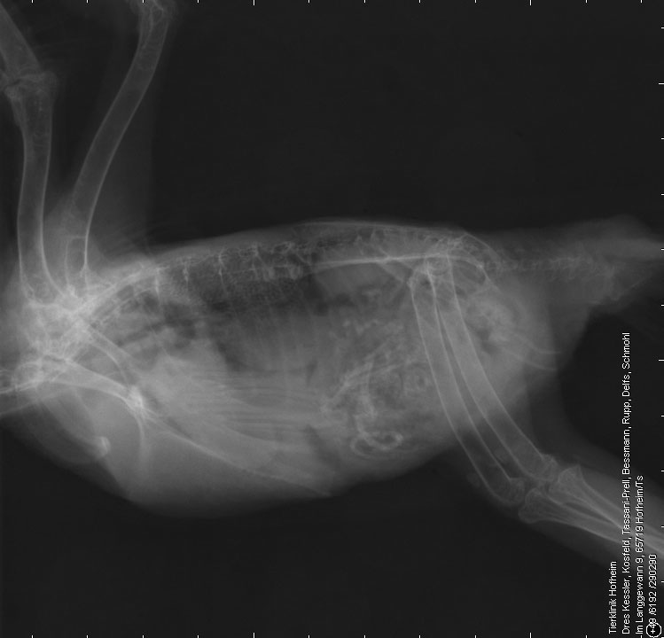
Barrel-chested thoracic confirmation. Normal body condition. Expiratory view with reduced thoracic volume on the lateral radiograph, good inspiratory ventrodorsal (VD) view. The 2nd sternebra is shorter with a flat caudal endplate. Mild step formation between 2nd and 3rd sternebae.

Spondylosis arising from the endplates of C6/7. Small amount of new bone formation at caudal aspect humeral heads.

The diaphragm is intact. The crurae are crossing T9 (expiratory view).

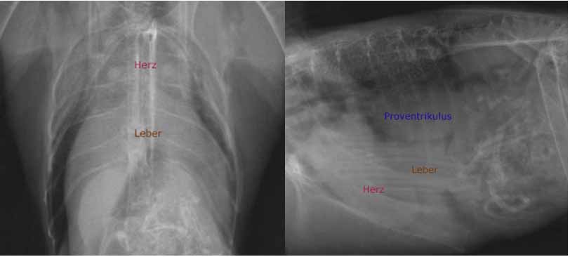
The liver is of homogenous soft tissue opacity. The stomach contains food material. On the lateral radiograph the neck is in a slightly flexed position. Larynogpharynx and larynx contain air. The cervical portion of the trachea is slightly narrower than the larynx, however overall it is wide. At the level of C6 the trachea shows an abrupt narrowing and runs dorsally resulting in an S-shaped kink.

Caudal to the kink the trachea has a reduced diameter, of approximately half the height of the larynx.
The intrathoracic portion of the trachea is barely visible and of soft tissue opacity.
At the level of the bifurcation the trachea as well as the main stem bronchi are not visible due to collapse.

At the level of C6, dorsal to the S-shaped tracheal kink, a crescent-shaped, clearly delineated gas accumulation is visible.

On the lateral radiograph the cardiac silhouette appears subjectively large, occupying 4 intercostal spaces and approximately 90% of the thoracic height. On the VD radiograph the cardiac silhouette is unremarkable. It occupies approximately 60% of the thoracic width.

No pathologic bulges are present on either view. The lung vessels are within normal limits. On the lateral radiograph the lung shows an overall increased opacity with a reticular pattern. Cranial to the cardiac silhouette a wing-like gas structure is visible extending from the cardiac silhouette into the caudal cervical area to C6. Centrally it contains slightly unsharp, fish bone like, soft tissue dens bands. On the VD radiograph the lung is within normal limits. The wing-like structure is not visible.

Radiographic diagnoses

  • Collapse intrathoracic portion of the trachea
  • Collapse of the main stem bronchi
  • Kinking caudal cervical trachea
  • Wing-like gas opaque structure cranial to the cardiac silhouette extending into the neck
  • Crescent shaped gas opaque structure dorsal to trachea
  • Interstitial lung pattern lateral radiograph
  • Spondylosis C6/7
  • Malformation 2nd sternebrae with step formation
  • Bilateral mild osteoarthritis shoulder joint

Discussion

The wing-like gas opaque structure cranial to the cardiac silhouette represent unilateral or bilaterally herniated cranial lung lobe(s). Lung lobe herniation is defined as the protrusion of lung parenchyma beyond the level of the thoracic boundaries. It is described to occur as a sequel to chronic respiratory distress and is strongly associated with collapse of the intra-thoracic trachea and major bronchi. Kinking of the extra-thoracic part of the trachea occurs in up to 1/3 of dogs.

The crescent shaped gas opaque structure dorsal to the trachea is most likely the gas filled oesophagus. Aerophagia could very well be explained by respiratory distress due to the tracheal and bronchial collapse. Most likely gas had been swallowed during acquisition of the radiograph.

The interstitial lung pattern is due to lack of lung aeration due to the expiratory nature of the lateral radiograph. The subjectively large cardiac silhouette on the lateral radiograph is due to the barrel-chested body confirmation and therefore the reduced thoracic height as well as the reduced thoracic volume due to expiration.

The malformation of the 2nd sternebrae is most likely due to an old trauma, differential diagnosis is a congenital malformation.

References

  • Dynamic Cervical Lung Hernia in a Dog with Chronic Airway Disease. Coleman et al. J. Vet. Int. Med. 2005
  • Cervical lung lobe herniation in dogs identified by fluoroscopy. Nafe et al. Can. Vet. J. 2013
  • Intermittend cranial lung herniation in two dogs. Guglielmini et al. Vet. Radiol. Ultraound 2007
Read More

Rottweiler, 19 months

Heart murmour, exercise intolerance

Images courtesy of Tierklinik Hofheim. Dres Kessler, Kosfeld, Tassani-Prell, Bessmann, Rupp, Delfs, Schmohl, von Klopmann

Diagnosis

Description

Expiratory radiographs.

The bony structures are within normal limits.

The diaphragmatic outline is not clearly visible ventrally as it is silhouetting with the cardiac silhouette. Steep orientation of the diaphragm.

The liver extends beyond the costal arch and the caudo-ventral liver margin is not clearly delineated.

The cardiac silhouette is enlarged. It occupies 4 intercostal spaces and 90% of the thoracic height on the lateral radiograph and there is buldging of the cranial contour of the cardiac silhouette. Increased sternal contact is present.

On the dorsoventral (DV) radiograph the cardiac silhouette appears mainly broadened. At the level of the 7th rib it occupies almost 100% of the thoracic width. The tip of the cardiac silhouette is displaced to the left side. There is prominent buldging of the cardiac silhouette between 7 and 11 o’clock. At the 7 o’clock the cardiac silhouette becomes slightly smaller; however, a convex bulging is still visible up to the level of the tip of the cardiac silhouette at 5 o’clock. The trachea runs parallel to the spine (lateral radiograph).

No splitting of the main stem bronchi is visible on the lateral view and no widening of the space between the two main stem bronchi is evident on the DV view.

The caudal vena cava (CVC) has an increased diameter and measures approximately 1.3times the height of the vertebral bodies.

The course of the CVC is normal, extending from a cranioventral in a caudodorsal direction.
The lung shows a mildly increased opacity with fine reticular pattern.

The vessel size is unremarkable.

A sail-like, soft tissue opaque structure is visible on the DV radiograph paralleling the left side of the cardiac contour.

Radiographic diagnoses

  • Severe, right sided cardiomegaly with bulging of the cardiac silhouette between 7–11 o’clock and to a lesser degree between 5–7 o’clock
  • Increased diameter of the CVC
  • Suspicion of hepatomegaly
  • Sail-like structure left hemithorax

Discussion

The buldging of the cardiac silhouette between 7-11 o’clock is compatible with an enlarged right atrium and right ventricle.

However, the severe dilation cannot be traced to the tip of the cardiac silhouette but instead terminates abruptly at 7 o’clock.

This makes it more likely that the bulge represents the severely enlarged right atrium. The bulge between 5-7 o’clock is compatible with an enlarged right ventricle which is less pronounced than the enlargement of the right atrium.

There are no signs for an enlarged main pulmonary artery.

The elevation of the trachea points towards an enlargement of the left side of the heart.
However, no splitting or bowing of the main stem bronchi is present and tenting of the LA is absent. Thus the left atrium is not enlarged.

Additionally, the caudal contour of cardiac silhouette is not straight or abnormally upright and there is no bulging between 12 -2 o’clock.

The normal course of the CVC speaks against a significant enlargement of the left ventricle, because this would lead to an elevation of the CVC.

On the DV view no bulging is visible along the left contour of the cardiac silhouette. Therefore, an enlargement of the left atrium and ventricle is unlikely.

The increased diameter of the CVC points towards the presence of a right-sided backward failure. Hepatomegaly would tie in with this finding and would be caused by hepatic congestion. Overall findings are compatible with an enlarged right atrium with right heart failure with hepatic congestion and enlargement of the CVC.

The young age of the dog makes a congenital disease most likely.

The sail like structure at the left side of the cardiac silhouette represents the thymic remnant, which is likely displaced by the enlarged heart and therefore better visible than expected in an 18 months old dog.

Differential diagnoses for right sided enlargement include:

  • Tricuspid valve dysplasia
  • Persistent artrio-ventriclar (AV) canal
  • Atrial septal defect
  • Cor triatrium dexter
  • Pulmonic stenosis

Tricuspide valve dysplasia is a possibility.
Due to the valvular insufficiency, regurgitant blood enters the right atrium during systole leading to atrial enlargement and possible right heart failure.

Persistent AV canal is possible, especially the complex form with a connection between all 4 chambers, which leads to an enlargement of right atrium and ventricle.

For a cor triatrium dexter the CVC is not big enough it cannot be eliminated from the list of differential diagnoses.

An atrial septal defect does usually not cause radiographic changes. Shunting of the blood usually occurs during diastole with blood entering the right ventricle, bypassing the right atrium. During systole the blood enters the lung. The resulting volume overload of the lung can lead to pulmonary hypertension with subsequent concentric hypertrophy of the R ventricular wall. A severe enlargement of the right atrium, as visible in this case, would be unlikely.

Pulmonic stenosis (PS) is unlikely due to the lack of an enlarged main pulmonary artery and normal sized lung vasculature (PS is associated with hypoperfusion).

Recommendations

Check the echogradiography for the final diagnosis.

Read More

Crossbreed dog Lucky, 9 years

Swallowing problems, respiratory distress

Images courtesy of Tierklinik Hofheim. Dres Kessler, Kosfeld, Tassani-Prell, Bessmann, Rupp, Delfs, Schmohl, von Klopmann

Diagnosis

Description

Good body condition. Inspiratory radiograph.

Mild new bone formation visible ventral to T4/5 and T12/13 (spondylosis). Small osteophyte visible at the infraglenoid tubercles of one scapula. Abdominal detail within normal limits.

There is mild convex bulging of the ventral border of the cranial mediastinum. Overall the opacity at the level of the cranial mediastinum appears increased. The trachea runs almost parallel to the thoracic spine with a mild dorsal buldging at the level of the 3rd to 4th intercostal space. Dorsal to the 2nd sternebra a poorly delineated soft tissue opaque structure is visible.

The lung shows an overall, severely increased opacity. Between the cardiac silhouette and the diaphragm it is of almost soft tissue opacity, however the diaphragmatic outline can still be identified. On close inspection also fine white lines can also be seen in the affected areas; they represent vessels. The remaining lung shows more reticular changes. Throughout the entire lung field multiple, soft tissue nodules are visible.

Radiographic diagnoses

  • Severe interstitial pattern with multiple small soft tissue nodules (reticulonodular)
  • Convexity of the cranial mediastinum
  • Soft tissue dens structure dorsal to the 2nd sternebra
  • Spondylosis
  • Mild osteoarthrosis shoulder

Discussion

The lung changes are highly suspicious for a neoplasia (primary versus secondary).

The changes of the cranial mediastinum are suspicious for a cranial mediastinal mass. Differential diagnoses include enlarged cranial mediastinal lymphnodes, mass arising from the oesophagus (neoplasia, granuloma) as well as mediastinal haemorrhage, abcesses, granuloma or inflammation (for example due to perforation of the oesophagus, nocardiosis).

The soft tissue structure dorsal to the 2nd sternebra is most likely an enlarged sternal lymphnode.

The severe opacification caudodorsal to the cardiac silhouette can be due to superimposition of severely altered lung parenchyma. As only a single lateral radiograph is available a caudal mediastinal mass, for example arising from the oesophagus, a hematoma, an abcess or a granuloma cannot be excluded. A second radiograph would be required to assess this change further.

In combination with the lung changes a neoplasia with metastasis to the sternal and mediastinal lymphnodes appears the most likely differential diagnosis.

Recommendations

  • Left lateral and ventrodorsal radiograph
  • And/or Computed tomography of the lung

Outcome

Computed tomography of the thorax confirmed the severe interstitial lung pattern with multiple soft tissue nodules. The sternal as well as the cranial mediastinal lymphnodes were severely enlarged.

Caudal to the heart no other mass lesions were visible, therefore the radiographic changes are most likely due to superimposition of the lung changes.

Lucky had a carcinoma of the larynx with metastasis to the lung and lymphnodes.

A heavy interstitial lung pattern can mimic an alveolar lung pattern. Don’t get fooled by the first impression. On close inspection there are usually still some vessels visible.

Read More

Kestrel

Found, no history provided

Images courtesy of Tierklinik Hofheim. Dres Kessler, Kosfeld, Tassani-Prell, Bessmann, Rupp, Delfs, Schmohl, von Klopmann

Diagnosis

Description

The left scapula shows a fracture line at the level of the scapular neck with craniomedial displacement of the fragment. The left clavicle also shows a fracture line. The coracoid is intact. Soft tissue swelling is present surrounding the left shoulder joint. On the lateral radiographs the fracture lines are not clearly visible due to superimposition.

On both views the silhouette of the liver is severely enlarged. On the ventrodorsal radiograph the thoracic and abdominal air sacs are only partially visible lateral to the enlarged liver. On the lateral radiograph an ovoid, soft tissue opaque structure is visible centrally in the coelomic cavity, representing the dorsally displaced proventriculus.

Radiographic diagnoses

  • Fracture of the left scapula and clavicle
  • Hepatomegaly

Discussion

The most likely cause for hepatomegaly is hepatic lipidosis. Differential diagnoses include infectious hepatitis and neoplasia.

Evaluation of the fracture age is difficult. The fracture ends appear blunted, however there is soft tissue swelling surrounding the left shoulder joint. It can be assumed that these are recent fractures with the soft tissue swelling representing a hematoma. The rounded appearance of the fracture margins can be artificial due to the small size of the object leading to slight blurring of small structures.

Read More
WordPress Cookie Notice by Real Cookie Banner