
Rottweiler, 19 months
Which imaging plane is shown in the videos? What conclusion can be drawn from the doppler image? What’s your diagnosis?
Diagnosis
Echocardiographic report
Video 1
Right parasternal long-axis 4 chamber view
The right atrium is severely enlarged. Additionally a mild enlargement of the right ventricle is evident.
Video 2
Left parasternal caudal/ apical 4 chamber view
Doppler imaging of the blood flow through the tricuspid valve. The tricuspid valve is not closing completely. Severe regurgitation is present during systole.
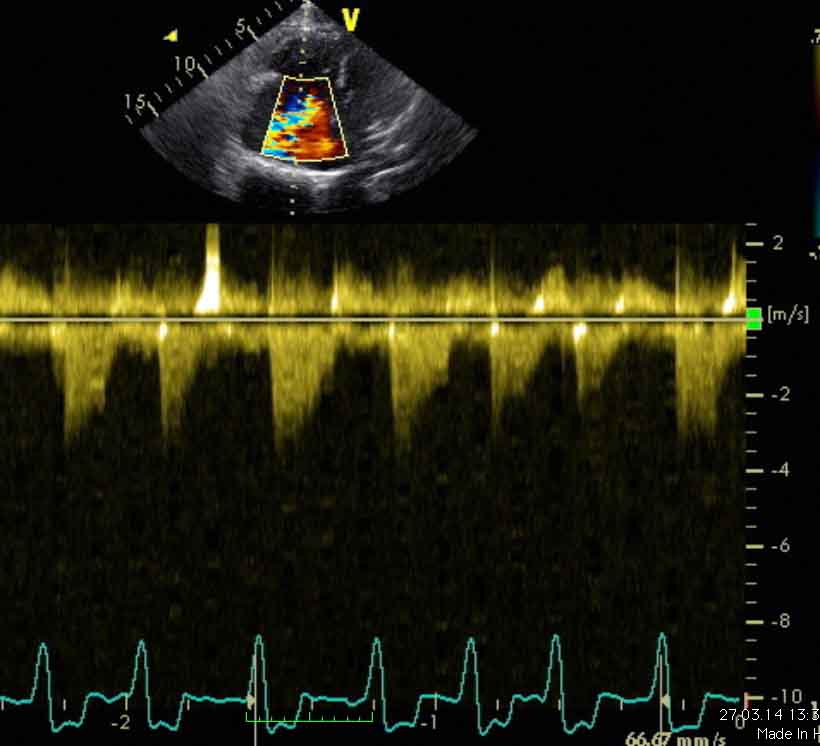
Image
Same position of the probe as in video 2
Continuous wave Doppler demonstrates the regurgitatant flow into the right atrium during systole. The image of the flow profile is suboptimal but the maximum velocity is approximately 4m/second.
The pressure in the main pulmonary artery (MPA) during systole can be calculated from the velocity of the regurgitant flow using the Bernoulli equation (p = 4 (V2)). With a maximum velocity of 4m/sec the pressure in the MPA is 64 mmHG.
The normal pressure in the MPA during systole is approx. 20 mmHg. The increased pressure in this case is indicative of pulmonary hypertension.
Additionally, the ECG shows an irregular rhythm and no visible P-waves. Thus, atrial fibrillations are present.
Echocardiographic diagnosis
Severe, decompensated tricuspid insufficiency due to tricuspid valve dysplasia.
X-rays can be found under the patient with the same name: