Portfolio CT

French Bulldog, Freya, 10 years

Hematochezia and suspected rectal neoplasia
Heart base mass noted on CT

CT dated Mai 2025, with a mass noted at the base of the heart, which was an incidental finding and asymptomatic at the time of imaging.

Diagnosis

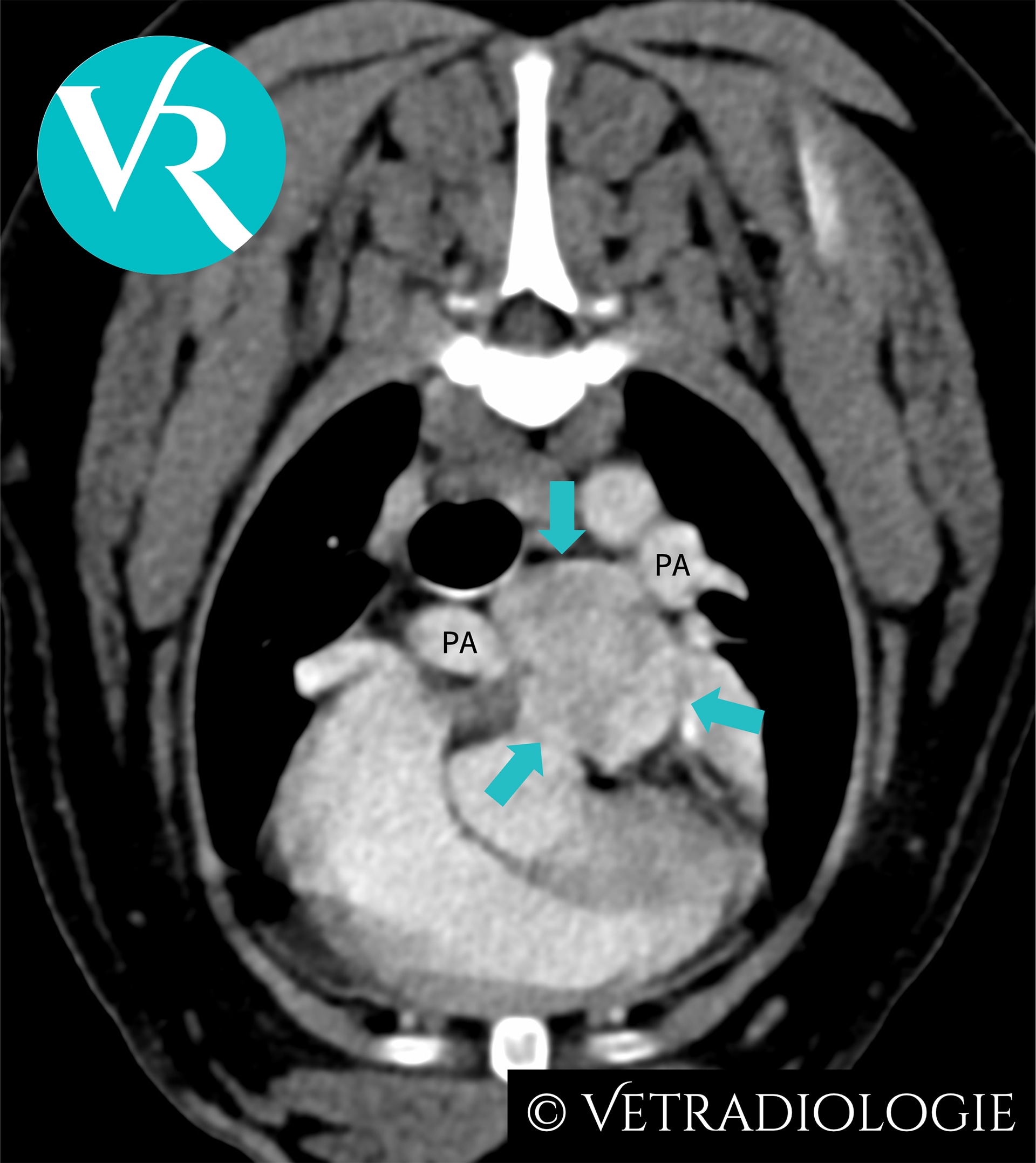
Freya’s CT Study: Note the well circumscribed, ovoid, heterogeneously contrast enhancing soft tissue mass (arrows) at the level of the main pulmonary arterial bifurcation.

CT-Findings

There is a well circumscribed, ovoid, hypervascular soft tissue mass (approximately 2.6cm in maximum diameter) located at the level of the main pulmonary arterial bifurcation, exhibiting heterogeneous contrast enhancement.

CT-Diagnosis

  • Heart base tumor

Discussion

Location and morphology are characteristic for a chemodectoma originating from the pulmonary glomus. A chemodectoma is a type of neuroendocrine tumor (NET) arising from chemoreceptor cells at the main pulmonary trunk. These chemoreceptors are organized into glomera and functionally assigned to the paraganglia. Tumors of these circulatory regulatory structures are referred to as chemodectomas or paragangliomas, and are reported more commonly in dogs than in cats.

Chemodectomas are typically benign, non-functional, and exhibit a low metastatic rate, although they may demonstrate locally invasive growth. Due to their slow growth, they frequently remain asymptomatic and—as in Freya—are often incidental findings. However, they may lead to clinical signs such as syncope, exercise intolerance, or sudden cardiac death.

In addition to the pulmonary glomus, chemodectomas/paragangliomas may also arise from other glomera such as from the aortic body glomus at the aortic root or carotid glomus at the bifurcation of the carotid artery.

The chemoreceptors of the glomera respond physiologically to reduced partial oxygen pressure and decreased blood pH. Chronic stimulation due to hypoxemia and respiratory acidosis, such as occurs in the context of the brachycephalic airway syndrome, has been discussed as a possible cause of the increased prevalence of chemodectomas in brachycephalic dogs

References

  • Buscaglia NA, Johnson PJ. What Is Your Diagnosis? J Am Vet Med Assoc. 2019;254(4):467-469. doi:10.2460/javma.254.4.467.
  • Crawford-Jennings MI, Chavez LD, Loessberg ER, Carvallo-Chaigneau FR. Aortic body tumor with intracardiac metastasis in a dog. J Vet Diagn Invest. 2025 Mar;37(2):345-348. doi: 10.1177/10406387241304438.
  • Fife W, Mattoon J, Drost WT, Groppe D, Wellman M. Imaging features of a presumed carotid body tumor in a dog. Vet Radiol Ultrasound. 2003;44(3):322-325. doi:10.1111/j.1740-8261.2003.tb00463.
  • Kromhout K, Gielen I, De Cock HE, et al. Magnetic resonance and computed tomography imaging of a carotid body tumor in a dog. Acta Vet Scand. 2012;54:24. doi:10.1186/1751-0147-54-24.
  • Mai W, Seiler GS, Lindl-Bylicki BJ, et al. CT and MRI features of carotid body paragangliomas in 16 dogs. Vet Radiol Ultrasound. 2015;56:374-383. doi:10.1111/vru.12251.
  • Obradovich JE, Withrow SJ, Powers BE, Walshaw R. Carotid body tumors in the dog. Eleven cases (1978–1988). J Vet Intern Med. 1992;6(2):96-101. doi:10.1111/j.1939-1676.1992.tb03158.
  • Wess G. Kardiale und perikardiale Tumoren. In: Kessler M, Hrsg. Kleintieronkologie. 4., vollständig überarbeitete Auflage. Stuttgart: Thieme; 2022:600f.
Read More

French Bulldog, Sammy, 9 years

Head tilt, right-sided Horner’s syndrome, right-sided facial discomfort

CT dated September 2021. Presented with 10d history of cough, dyspnea and nasal discharge. Acute right-sided facial paralysis and lethargy.

Diagnosis

Sammy’s CT-Study: Infiltrative soft tissue mass (turquoise arrows) exhibiting marked heterogeneous contrast enhancement and invasion of the cranial vault (yellow arrow) along the plane of the right internal carotid artery (red arrow).

CT-Findings

Right-sided retropharyngeal soft tissue mass located medial to the right mandibular salivary gland in the region of the common carotid arterial bifurcation, with craniodorsal mass extension towards the cranial vault. There is associated invasion of the digastric and medial pterygoid muscles, and osteolysis of the right temporal bone (including petrous, tympanic and squamous parts). There is extension of the lesion into the caudal fossa via moth-eaten and permeative osteolysis of the squamous part of the right temporal bone, with the intracranial mass component reaching and conforming to the tentorium cerebelli ventrally.

Caudally, the lesion extends along the right common carotid artery to the approximate level of the thyroid gland.
The lesion exhibits marked, heterogeneous contrast enhancement.

CT-Diagnosis

  • Aggressive, invasive soft tissue mass originating near the bifurcation of the right common carotid artery.

Discussion

The imaging features of the mass are consistent with a carotid body tumor/glomus caroticum tumor. This subtype of paraganglioma belongs to the group of neuroendocrine tumors (NETs).

Brachycephalic dog breeds are predisposed.

Carotid body tumors arise from the neuroendocrine chemoreceptors of the carotid glomus, located at the bifurcation of the common carotid artery. Similar clusters of chemoreceptors (glomera) exist in the wall of the aorta (aortic body glomus) and the pulmonary trunk (pulmonary glomus), where tumor development (chemodectomas, glomus aorticum tumors) has likewise been described.

As circulatory regulatory structures, the chemoreceptors of the glomera respond to reduced oxygen partial pressure and to changes in blood pH.

Chronic hypoxia and respiratory acidosis associated with brachycephalic obstructive airway syndrome (BOAS) lead to persistent stimulation of these chemoreceptors. It is assumed that this—either alone or in combination with a genetic component—accounts for the described breed predisposition to the development of carotid body tumors and chemodectomas.

The clinical presentation varies depending on tumor size and the extent of involvement of adjacent structures. Potential symptoms include regional discomfort, head tilt, coughing, dyspnea, Horner’s syndrome, or dysphagia.

On CT, carotid body tumors appear as highly vascularized, locally invasive masses with marked, heterogeneous contrast enhancement.

References

  • Buscaglia NA, Johnson PJ. What Is Your Diagnosis? J Am Vet Med Assoc. 2019;254(4):467-469. doi:10.2460/javma.254.4.467.
  • Crawford-Jennings MI, Chavez LD, Loessberg ER, Carvallo-Chaigneau FR. Aortic body tumor with intracardiac metastasis in a dog. J Vet Diagn Invest. 2025 Mar;37(2):345-348. doi: 10.1177/10406387241304438.
  • Fife W, Mattoon J, Drost WT, Groppe D, Wellman M. Imaging features of a presumed carotid body tumor in a dog. Vet Radiol Ultrasound. 2003;44(3):322-325. doi:10.1111/j.1740-8261.2003.tb00463.
  • Kromhout K, Gielen I, De Cock HE, et al. Magnetic resonance and computed tomography imaging of a carotid body tumor in a dog. Acta Vet Scand. 2012;54:24. doi:10.1186/1751-0147-54-24.
  • Mai W, Seiler GS, Lindl-Bylicki BJ, et al. CT and MRI features of carotid body paragangliomas in 16 dogs. Vet Radiol Ultrasound. 2015;56:374-383. doi:10.1111/vru.12251.
  • Obradovich JE, Withrow SJ, Powers BE, Walshaw R. Carotid body tumors in the dog. Eleven cases (1978–1988). J Vet Intern Med. 1992;6(2):96-101. doi:10.1111/j.1939-1676.1992.tb03158.
  • Wess G. Kardiale und perikardiale Tumoren. In: Kessler M, Hrsg. Kleintieronkologie. 4., vollständig überarbeitete Auflage. Stuttgart: Thieme; 2022:600f.
Read More

Labrador Retriever Bella, 9 years

Progressive swallowing problems, especially of larger items

Post-contrast sequence

Pre-contrast sequence

With kind permission Dr. G. Viefhues, Small Animal Clinic Ahlen

Diagnosis

Description

The atlanto-occipital and atlanto-axial junctions are surrounded by a cavitating, space occupying lesion which is hypoattenuating to the surrounding soft tissue. The density of the individual cavities is 11-13 HU (density of the neighboring muscles is approx. 80 HU). Another space occupying lesion is located ventral to the M. longus capitis and appears separate from the other masses. It is approx. 3.8cm long, 2.5cm high, 4.1cm wide and results in severe displacement of the pharyngeal roof so that the caudal nasopharynx is completely obliterated. The occipital and temporal bones as well as the atlas (C1) and cranial aspect of the axis (C2) including the dens show a large number of well circumscribed, lytic defects. The space occupying lesion extends into the right caudal cranial fossa which leads to mild displacement and compression of the cerebellar vermis. Brain stem and spinal cord at C1 and C2 are also compressed; mostly from ventral and the sides. Severe, heterogenous and foamy contrast enhancement of the compressing structures in the spinal canal is evident. The space occupying lesions do not contrast enhance.

CT diagnosis

  • Soft tissue neoplasia atlanto-occipital and atlanto-axial junction with bone destruction and compression of cerebellum, pons and spinal cord

Discussion

The changes are suggestive of a myxosarcoma. Differential diagnoses include all other soft tissue tumours; however, the extreme hypodensity of all masses is highly suggestive of a myxosarcoma. The tumor is most likely arising from the atlanto-occipital and atlanto-axial joints, which are continuous which each other. The described masses are therefore most likely connected and part of one tumor mass.

Outcome

Bella was euthanized. The histopathological report confirmed the diagnosis of myxosarcoma.

Read More

Small Münsterländer Samson, 7 years

For 4 weeks: swelling lateral to the L eye, no improvement on antibiotics and pain killers: trauma cannot be ruled out.

With kind permission Dr. G. Viefhues, Small Animal Clinic Ahlen

Diagnosis

Description

An approx. 3cm large, mineral dense mass with a granular appearance extends from the zygomatic process of the left maxilla to the zygomatic arch. The mass protrudes slightly into the orbital cavity and causes mild deformity of the globe. The underlying cortex has a slightly roughened surface and shows small, fine osteolytic regions. An extension of the mass into the bone marrow is not evident.

CT diagnosis

  • Bony mass left zygomatic arch

Discussion

The appearance of the tumor is typical for a multilobular tumor of the bone. Other names are osteochonrosarcoma and chondrosarcoma rodens.

Read More

GSD Dexter, 8 weeks

Mucohemorrhagic nasal discharge, painful swelling left maxilla

Images courtesy of Kleintierklinik Greven. Dres. Böhmer, Cordes, Möller and Wienker

Diagnosis

Description

Large expansile, cystic lesions are visible in the right maxilla surrounding the crowns of the unerupted permanent 4th premolar (PM4), as well as of the 1st and 2nd molar (M1 and M2). The oral part of the cortex is partially disrupted at the level of M1 and M2. No connection to the nasal cavity is visible. Large expansile, cystic lesions are also surrounding the unerupted permanent mandibular incisor teeth.

After contrast medium administration moderate, heterogenous contrast uptake is visible within the cystic lesion in the right maxilla and in the incisor part of both mandibles.

A severe reduction in the number of nasal turbinates is present in both nasal cavities. The remaining turbinates appear plump. Material isodense to soft tissue is present between the remaining turbinates. The nasopharyngeal meatus is narrowed and contains a moderate amount of material isodens to soft tissue, which circumferentially occupies approximately 60% of its diameter.

Severe enlargement of the mandibular and the medial retropharyngeal lymph nodes is present bilaterally (left > right).

Radiographic diagnoses

  • Expansile, osteolytic bone lesions associated with the crowns of the unerupted permanent teeth (P4, M1 and M2 right maxilla, incisor teeth both mandibles)
  • Bilateral reduction in nasal turbinates
  • Soft tissue material between remaining nasal turbinates
  • Lympahdenomegaly mandibular and medial retropharyngeal lymphnodes

Discussion

The changes are suggestive of dentigerous cysts. As differential diagnosis odontogenic keratocysts should be considered. However, odontogenic kertocysts are rare in dogs. Differentiation of the various odontogenic cysts required histopathology examination of the wall of the cysts.

The changes in the nasal cavity are suggestive of a congenital hypoplasia of the nasal conchae with secondary bacterial infection. As a differential diagnosis to a simple conchael hypoplasia a ciliary dyskinesia with or with situs inversus (Kartagener Syndrom) should be considered. A chronic bacterial rhinitis with secondary destruction of the nasal conchae is less likely due to the young age of the dog.

Read More

Irish Wolfhound Bailey, 1 year

Initially left sided, purulent nasal discharge, later bilateral nasal discharge

CT images from July 2014. At that time unilateral nasal discharge was present.

CT images from March 2015. This time bilateral nasal discharge was present.

Images courtesy of Kleintierklinik Greven. Dres. Böhmer, Cordes, Möller and Wienker

Diagnosis

Description

Severe reduction in size and number of the turbinates in both nasal cavities. Between the remaining turbinates a mild to moderate amount of soft tissue dense material is present. Within the left frontal sinus soft tissue dense material with a fluid level is present (images flipped horizontally, patient was placed in dorsal recumbency during image acquisition). The mucosal lining of the left frontal sinus is thickened. Findings are almost identical in both CT studies.

Radiographic diagnoses

  • Bilateral reduction in number and size of nasal turbinates
  • Soft tissue dense material between the turbinates
  • Sinusitis with fluid accumulation left frontal sinus

Discussion

Changes are compatible with rhinitis- bronchopneumonia syndrome of the Irish Wolfhound. The etiology is unclear. Immune mediated or primary ciliary defects are discussed. A hereditary background is assumed. Affected dogs show transient or persistent mucoid to mucopurulent nasal discharge of variable degree from birth . Concurrent bronchopneumonia can occur (most likely due to the ciliar dysfunction leading to an increased susceptibility for infection).

No progression of the changes is seen over the one year period. No granuloma is visible in the frontal sinus. The paranasal bones are unremarkable, showing no permeative osteolysis. Therefore a fungal rhinitis is less likely. In addition the dog is very young making a fungal rhinitis unlikely as well.

Recommendations

None.

Normal CT study nasal cavity

A reduction in the number of turbinates is not always related to a destructive rhinitis. Consider congenital hypoplasia predisposing the nasal cavity to infection, especially in young dogs.

Read More
WordPress Cookie Notice by Real Cookie Banner