Wood pigeon


Diagnosis
Description
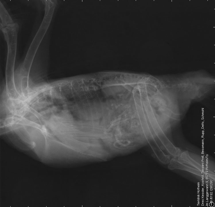
Radius, ulna and tibiotarsal bones show multiple expansile lesions with central osteolysis and thinning of the overlying cortex. Adjacent to the osteolytic areas the bones are sclerotic. A complete fracture through the osteolytic area in the mid diaphysis of the right radius is present. The left ulna shows a fracture at the level of the osteolytic area in the distal metaphysis resulting in severe shortening of the bone and cranial displacement of the radius. Another complete fracture is visible in the distal diaphysis of the right tibiotarsal bone. Soft tissue swelling is present surrounding the fracture sites.
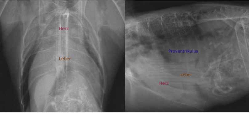
On the lateral radiograph the lung shows a generalised increase in opacity; the liver is enlarged.
Radiographic diagnoses
- Multifocal, expansile bone lesion
- Multiple pathological fractures
- Luxation of the left radius due to a pathologic compression fracture of the left ulna resulting in shortening of the ulna
- Pulmonopathy
- Hepatomegaly
Discussion
The changes are compatible with mycobacteriosis. Due to the close bond between the respiratory system and the pneumatised bone, infections often spread between the two organ systems. Pulmonary lesions commonly found in other species are, however, rare in birds.
Osteomyelitis of a different origins will have to be considered as a differential diagnosis.
The hepatomegaly could be caused by hepatic lipidosis, which is common in Wood pigeons. However, a connection between infection and hepatomegaly is also possible. Avian tuberculosis in domestic birds is primarily an intestinal and hepatic disease with dissemination to other organs including the lungs, air sacs, spleen, bone marrow, and skin. In case of infection of liver and spleen enlargement of the organ can occur.
Outcome
The pigeon was euthanized. Histo-pathologic examination confirmed granulomatous osteomyelitis due to mycobacteriosis.
In birds the lung opacity should always be assessed on the lateral radiograph. On ventrodorsal radiographs pseudo-opacification occurs due to superimposition of the pectoral muscles.